
EMMERGENCY:
(+237) 693 172 452 (+237) 693 172 453
IMMIGRATION:
(+237) 674 380 349
(+237) 693 172 452 (+237) 693 172 453
(+237) 674 380 349
DMMC provides Radiology and Medical Imaging services designed to support accurate diagnosis and effective treatment planning. Our department is equipped with modern imaging technologies and managed by qualified radiologists and technicians.
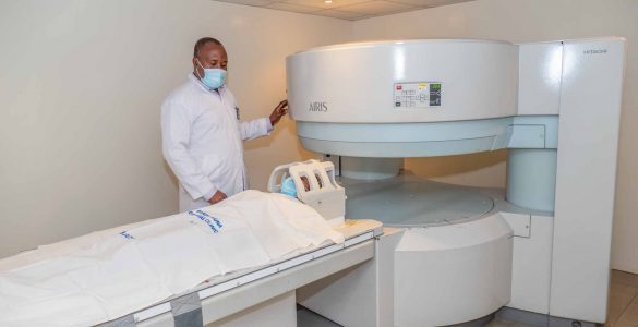
This service is made up of the following units:
Radiography, commonly known as X-ray, is the oldest imaging modality, discovered by German physicist Wilhelm Röntgen in 1895.
It works using ionizing radiation generated from an X-ray tube. When these rays pass through a body part and reach a photographic plate, they create an image of the internal structures.
A similar principle is used in mammography (breast examination), where softer X-ray beams are used. Screening mammography is performed even when no symptoms are present, for early detection of breast conditions.
Ultrasound imaging, also called sonography or echography, uses high-frequency sound waves (2 MHz – 10 MHz) to produce images of internal organs and tissues.
The sound waves pass through the body and are reflected back to create images used for diagnosis.
It is commonly used in:
A CT Scan combines multiple X-ray images taken from different angles and uses computer processing to create detailed cross-sectional images of bones, blood vessels, and soft tissues.
It provides more detailed information than standard X-rays and can be used to examine almost any part of the body.
CT scans may be performed:
In some cases, the radiologist may recommend a contrast agent to improve tissue visualization.
MRI is an advanced imaging technique that uses magnetic fields and radio waves to produce detailed images of soft tissues, organs, and bones.
It is commonly used for examining:
Unlike X-rays and CT scans, MRI does not use ionizing radiation.
Our team of experienced radiologists and radiology technicians is available to guide and support you throughout your imaging procedure and ensure accurate, high-quality diagnostic results.